Dr. Anne Royou
Research officer (DR2), CNRS
Cette adresse email est protégée contre les robots des spammeurs, vous devez activer Javascript pour la voir.
Website
Tel: +33(0)540006306
Bio
Following a bachelor degree in physiology and cell biology, Anne Royou did a postgraduate degree in molecular and cellular genetics at the Université Paris XI. She did her PhD thesis under the guidance of Dr. Roger Karess, at the Centre de Génétique Moléculaire in Gif-sur-Yvette, studying the role of non-muscle myosin II during development in Drosophila. Following her PhD, she joined Dr. William Sullivan’s lab at the University of California, Santa Cruz, as a post-doctoral fellow. There, she became interested in the mechanisms that preserve genome integrity during cell division. She obtained a CNRS permanent position in 2009, an ATIP/Avenir grant in 2010 and was recruited as a team leader at IECB in 2011. In 2013 she was awarded an ERC starting grant.
Summary
The mechanisms that safeguard cells against aneuploidy are of great interest as aneuploidy contributes to tumorigenesis. Using live imaging approaches, we have identified two novel mechanisms that permit the accurate transmission of chromosomes during cell division. The first mechanism involves the faithful segregation of damaged chromosomes. Our studies reveal that chromosome fragments segregate properly to opposite poles. This poleward motion is mediated through DNA tethers that connect the chromosome fragments. The second mechanism involves the coordination of chromosome segregation with cell cleavage. We found that cells can adapt to a four-fold increase in chromatid length by elongating transiently during anaphase. This mechanism ensures the clearance of chromatids from the cleavage plane at the appropriate time during cytokinesis, thus preserving genome integrity.
Activity report
Mitosis is the final stage of the cell cycle where a copy of the duplicated genome condensed into chromosomes is transmitted to each daughter cell. Failure to do so produces daughter cells with an inappropriate genome content, also called aneuploidy. The mechanisms that safeguard cells against aneuploidy are of great interest as aneuploidy contributes to tumorigenesis. Our group has identified two novel mechanisms that permit the accurate transmission of chromosomes during cell division. The first mechanism involves the faithful segregation of damaged chromosomes. The second mechanism coordinates chromosome segregation with cell cleavage.
Mechanism that permits faithful transmission of broken chromosomes
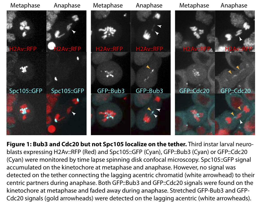
Correct transmission of the genetic material requires proper chromosome attachment to spindle microtubules. The centromere of the chromosome serves as an assembly site for a multi-protein structure called the kinetochore, the functional unit that binds the microtubules. The centromere is, thus, essential for proper chromosome segregation. However, we have recently identified a mechanism by which fragments of broken chromosomes lacking centromeres segregate properly. This is achieved through a novel chromatin conformation that tethers the acentric chromosome fragments to their centric partners. The efficient segregation of the broken chromosome fragments depends on the spindle assembly checkpoint component BubR1. BubR1 localizes on the kinetochore until anaphase onset and accumulates on the “tether”, the chromatin structure that attaches the chromosome fragments, throughout mitosis. To determine how BubR1 recruitment to the “tether” is regulated, we monitored dividing cells expressing BubR1 constructs lacking specific domains. We found that the conserved Bub3-Binding Domain and more specifically the Glutamate 481 (E481) are required for BubR1 localization on the tether. Since E481 is essential for BubR1 interaction with Bub3 and its localization on the kinetochore, we investigated whether Bub3 localizes to the tether and more generally whether the “tether” is an assembly site for other kinetochore proteins (Figure 1). We analysed the localization of five core kinetochore proteins and did not detect any signal on the “tether”. In contrast, Bub3 accumulated on the “tether” throughout mitosis. Bub3 and BubR1 were inter-dependent for their localization on the kinetochore and tether. In addition, we found that Cdc20, a key subunit of the APC/C, was recruited on the “tether” in a BubR1 and Bub3 dependent manner throughout mitosis (Figure 1). The expression of a BubR1 mutant lacking the KEN box, a motif required for Cdc20 binding to BubR1, exhibited severe defects in broken chromosome segregation and was associated with a lack of Cdc20 localization on the tether. To test if BubR1 inhibited locally the APC/C on the “tether”, we constructed a synthetic APC/C substrate. To do so, we fused the N terminal sequence of Cyclin B to GFP::Bub3 (DB-Bub3) and monitored its dynamics in the presence of broken chromosomes. We found that DB-Bub3 persisted on the “tether” during early anaphase in a BubR1 KEN box dependent manner. All together these results favour a model in which Bub3 recruits BubR1 on broken chromosomes, which, in turn, inhibits the APC/C locally by sequestering Cdc20 throughout mitosis. This local APC/C inhibition during anaphase preserves key components important for maintaining the broken fragments tethered.

Mechanism that coordinates chromosome segregation with cell cleavage
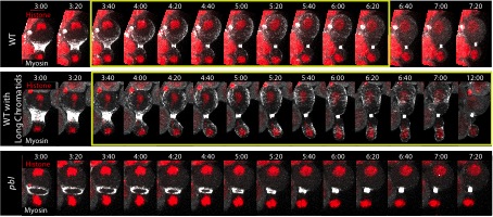
Chromosome segregation must be coordinated with cell division to ensure proper transmission of the genetic material into daughter cells. Our group identified a novel mechanism by which Drosophila neuronal stem cells coordinate chromosome segregation with cell division. Cells adapt to the presence of chromatid arms at the cleavage site by transiently, but dramatically, elongating during anaphase, thus increasing the rate at which the long chromatids clear the cleavage plane. This adaptive elongation depends on myosin activity and the Rho Guanine-nucleotide exchange factor, Pebble. To understand how the cell deforms when chromatids are present at the cleavage plane, we studied the dynamics of myosin during cytokinesis in cells with or without long chromosomes. Surprisingly, halfway through cytokinesis, myosin undergoes flux from the cytokinetic ring and invades the entire cortex. In control cells, myosin efflux is transient, as myosin dissociates from the cortex upon reformation of the nuclear envelope. However, cells with long chromatids exhibit an extended period of myosin efflux associated with a delay in nuclear envelope formation (NEF). During this prolonged myosin efflux, cortical myosin forms extra rings that deform the nascent daughter cells, permitting the clearance of chromatids from the cleavage plane. We have previously shown that adaptive cell elongation depends on Pebble. Consistently myosin does not undergo efflux in pebble mutant cells with or without long chromatids. Since Pebble accumulates in the nucleus at telophase at the time myosin dissociates from the cortex, we hypothesized that the translocation of Pebble into the nucleus induces myosin dissociation from the cortex via the inactivation of RhoA. Consistently, the expression of a pebble mutant lacking a nuclear location signal induces prolonged myosin efflux associated with cell elongation and extensive blebbing, even in the absence of long chromatid arms at the cleavage site. We propose that the coordination between chromatid segregation, myosin efflux and NEF allows adaptive cell elongation to clear chromatid arms from the cleavage plane before the completion of cytokinesis.

Figure 2: Myosin efflux during cytokinesis is prolonged in the presence of long chromatids and absent in pbl mutants.
Time-lapse images of wild type cells with normal chromosomes (top row), wild type cells carrying an abnormally long chromosome (second row), and cells mutant for the Rho-GEF Pebble (pbl, bottom row). The chromosomes are marked with H2Av::RFP (red) and myosin with Sqh::GFP (Gray). In control cell, half way through cytokinesis, myosin undergoes flux from the contractile ring and invades transiently the whole cortex (yellow rectangle) before dissociating from the cortex. This myosin efflux is prolonged during the segregation of long chromatids (yellow rectangle). No myosin efflux is detected in the pebble mutant. Time=min:second.
Selected publications
- Derive N*, Landmann C*, Montembault E, Claverie MC, Pierre-Elies P, Goutte-Gattat D, Founounou N, McCusker D and Royou A (2015) Bub3/BubR1-dependent sequestration of Cdc20Fizzy at DNA breaks facilitates the correct segregation of broken chromosomes. J Cell Biol. 211(3):517-32. * denotes equal contribution
- Jose M, Tollis S, Nair D, Mitteau R, Velours C, Massoni-Laporte A, Royou A, Sibarita JB, McCusker D (2015) A quantitative imaging-based screen reveals the exocyst as a network hub connecting endocytosis and exocytosis. Mol. Biol. Cell. 26(13):2519-34
- Kotadia, S*, Montembault, E*, Sullivan, W and Royou A (2012) Cell elongation is an adaptive response for clearing long chromatid arms from the cleavage plane. J Cell Biol. 199(5):745-53. * denotes equal contribution, # denotes corresponding author
- McCusker D*, Royou A, Velours C, Kellogg D* (2012) Cdk1-dependent control of membrane trafficking dynamics. Mol Biol Cell. Sep;23(17):3336-47. * denotes equal contribution.
- Royou, A., Gagou, M., Karess, R., D., Sullivan, W. (2010) BubR1 and Polo-coated DNA tethers facilitate the segregation of acentric chromatids. Cell 140(2) : 235-45.
- Royou, A., McCusker, D., Kellogg, D., Sullivan, W. (2008) Grapes(Chk1) prevents nuclear Cdk1 activation by delaying Cyclin B nuclear accumulation. J. Cell Biol. 183(1):63-75
- Riggs, B., Fasulo, B., Royou, A., Mische, S., Cao, J., Hays, TS., Sullivan, W. (2007) The concentration of Nuf, a Rab11 effector, at the microtubule-organizing center is cell cycle regulated, dynein-dependent, and coincides with furrow formation. Mol. Biol. Cell. 18(9) : 3313-22
- Chodagam, S., Royou, A., Whitfield, W., Karess, R., Raff, JW. (2005) The centrosomal protein CP190 regulates myosin function during early Drosophila development. Curr. Biol. 15 (14) : 1308-13
- Royou, A., Macias, H., Sullivan, W. (2005) The Drosophila Grp/Chk1 DNA damage checkpoint controls entry into anaphase. Curr. Biol. 15(4) :334-9
- Silva, E., Tiong, S., Pedersen, M., Homola, E., Royou, A. Fasulo, B. Siriaco, G. Campbell, S.D. (2004) ATM is required for telomere maintenance and chromosome stability during Drosophila development. Curr. Biol. 14(15) :1341-7
- Royou, A., Field, C., Sisson, J. Sullivan, W., Karess, R. (2004) Reassessing the role and dynamics of nonmuscle myosin II during furrow formation and cellularization in the early Drosophila embryo. Mol. Biol. Cell. 15: 838-50
- Royou, A., Sullivan, W., Karess, R. (2002) Cortical recruitment of non muscle myosin II in early syncytial Drosophila embryos: its role in nuclear axial expansion and its regulation by Cdc2 activity. J. Cell. Biol. 158(1):127-37
Research Team
- Dr. Anne Royou, Team leader (DR2, CNRS)
- Marie-Charlotte Claverie, Assistant Engineer (Université de Bordeaux)
- Dr. Emilie Montembault, Researcher (CR2, CNRS)
- Priscilla Pierre-Ellies, Assistant Engineer (ERC-STG-2012 NoAneuploidy)
- Lou Bouit, Assistant Engineer (ANR retour-postdoc)
- Dr. Damien Goutte-Gattat, Postdoctoral fellow (ERC-STG-2012 NoAneuploidy)
- Cedric Landmann, PhD student (RĂ©gion Aquitaine/CNRS)
- Enzo Camara, Master 2 student (CNRS)
- Camille Vachon, Master 2 student (CNRS)
- JĂ©rĂ´me Toutain, Visiting scientist (CHU Bordeaux/CNRS)
This team is part of the "Institut de Biochimie et Génétique Cellulaire" (IBGC), CNRS/Université Bordeaux (UMR5095)
|