| IECB at the cutting edge of ion mobility |

IECB at the cutting edge of ion mobility The Structural Biophysico-Chemistry platform is now equipped with a state-of-the-art drift tube ion mobility spectrometer: the first one available in plateform in Europe!
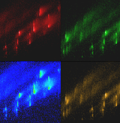
The ion mobility spectrometer (IMS) measures the motion speed of molecules or molecular complexes under a weak electric field. This precious data allows us to determine their precise conformation; which is the 3rd dimension it lacked when using only the mass spectrometry approach. Based on the notion that each molecular conformation has a specific time-of-flight (inversely proportional to the mobility), the IMS provides indications on the 3D-structure of a molecule. The detailed structure is then obtained with molecular modeling processes. These new datas are particularly interesting to determine the structure or the way biological molecules such as proteins or nucleic acids fold into well-defined conformations. It also is interesting regarding molecular complexes. Moreover IMS provides a better understanding of interactions and self-assembling mechanisms of supramolecular complexes such as G-quadruplex or foldamers, on which numerous IECB teams work on. This brand new IMS is installed on the mass spectrometry platform to which the research team of Dr. Valérie Gabelica, specialist of mass spectrometry of nucleic acids and supramolecular complexes, is closely link to. The IMS is available not only to the IECB research teams but also to the scientific European community via the COST BM1403 network. It was funded by the Aquitaine Regional Council (50%) and 50% by GIS IBISA, CNRS, Inserm, IECB and the ATIP-Avenir grant Dr Valérie Gabelica obtained when she joined the IECB in 2013.
*: IMS theory: the smaller or the more folded the molecule, the faster it moves in the drift tube, and vice versa for a wide broad molecule .
V. D’Atri, M. Porrini, F. Rosu, V. Gabelica, Special feature: tutorial. “Linking molecular models with ion mobility experiments. Illustration with a rigid nucleic acid structure”, J. Mass Spectrom. (2015), 50(5), 711-726 (issue cover). |
2, Rue Robert Escarpit - 33607 PESSAC - France
Tel. : +33 (5) 40 00 30 38 - Fax. : +33 (5) 40 00 30 68


